Scientists from Samara University and Samara State Medical University have patented the method they developed for the laser express-diagnostics of periodontitis the inflammatory disease of the gum tissue that can lead to tooth loss.
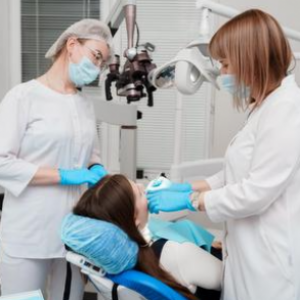
Using this method, the dentist can literally in a matter of seconds, by shining the laser on the questionable tooth, determine by using special equipment whether the patient has periodontitis, at what stage the disease is, or what is the probability of periodontitis in the near future. The prototype diagnostic system based on this method has already been tested in one of the dental clinics in Samara.
"Our joint research group, which includes scientists from Samara University and Samara State Medical University, received the patent of the Russian Federation for the developed method of diagnosing chronic periodontitis, based on the organic and mineral composition of tooth enamel. The patent for the invention officially certifies and confirms the priority of our development and the exclusive right to authorship, which will help us further introduce this technology more widely into dental practice both in our region and in the entire country, as well as develop this method of the express-diagnostics of periodontitis by designing new, more modern equipment samples", said Elena Timchenko, Associate Professor of Samara University’s Department of Laser and Biotechnical Systems, Deputy Scientific Director of the Interuniversity R&D Laboratory "Tissue Engineering".
Periodontitis is rather common dental disease. According to open sources, in the age group from 35 to 44 years, this pathology occurs in more than 80 % of Russians who seek help from a dentist. Periodontitis affects almost all the tissues surrounding the tooth, including those that connect the tooth to the jawbone, which ultimately, if left untreated, leads to tooth loss. Therefore, it is very important to detect this disease as early as possible.
The main factors of periodontitis are infections, weak immunity and poor oral hygiene. With chronic periodontitis at the initial stage, the patient may sometimes experience itching and soreness in the gums, bleeding when brushing teeth, and bad breath. However, these symptoms are also characteristic of other dental diseases, and in the earliest stages periodontitis can be completely asymptomatic and undetectable. The classic methods of diagnosing periodontitis involve X-rays and taking tests, but if the patient does not complain about anything, the doctor may skip the onset of the disease when examining the teeth.
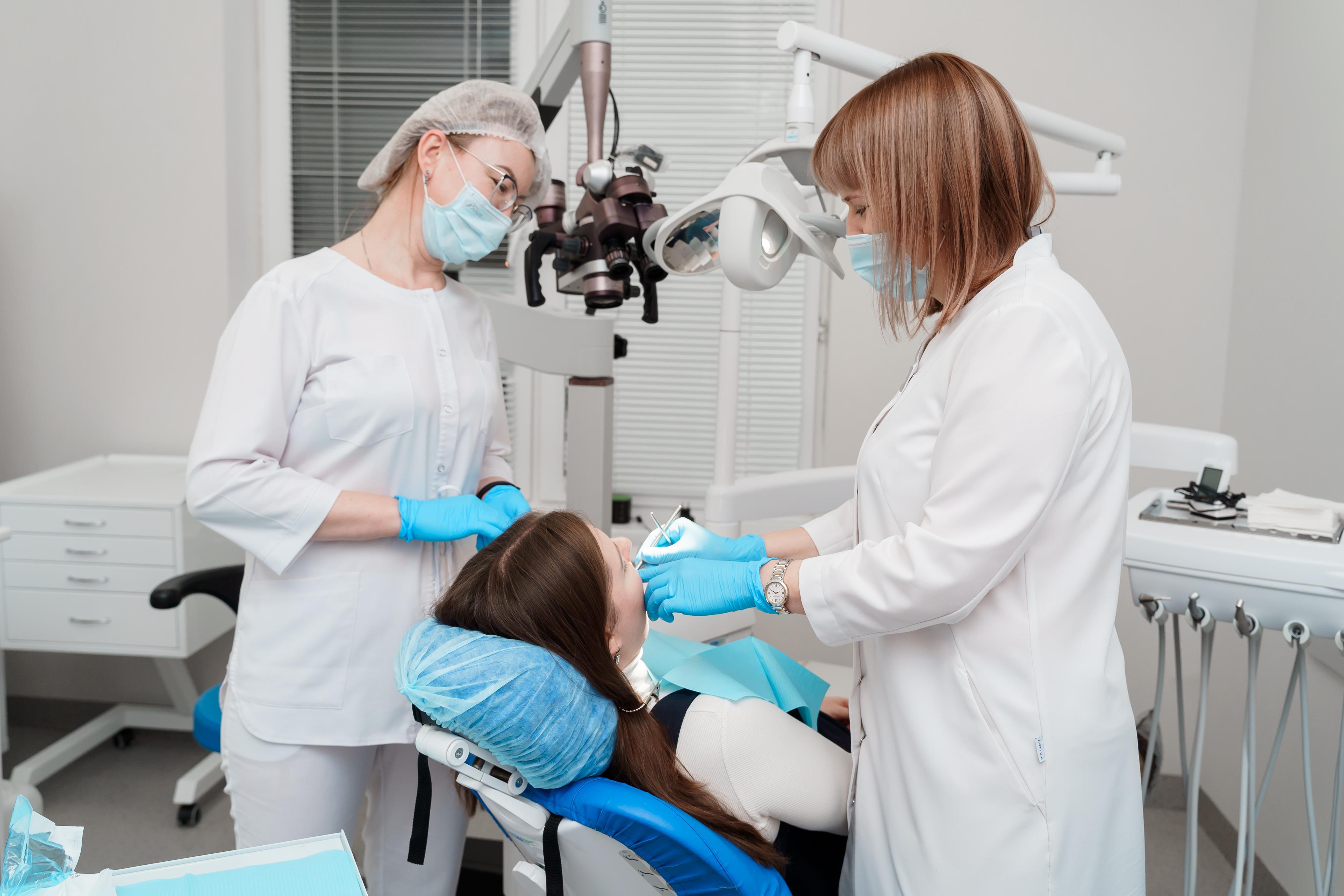
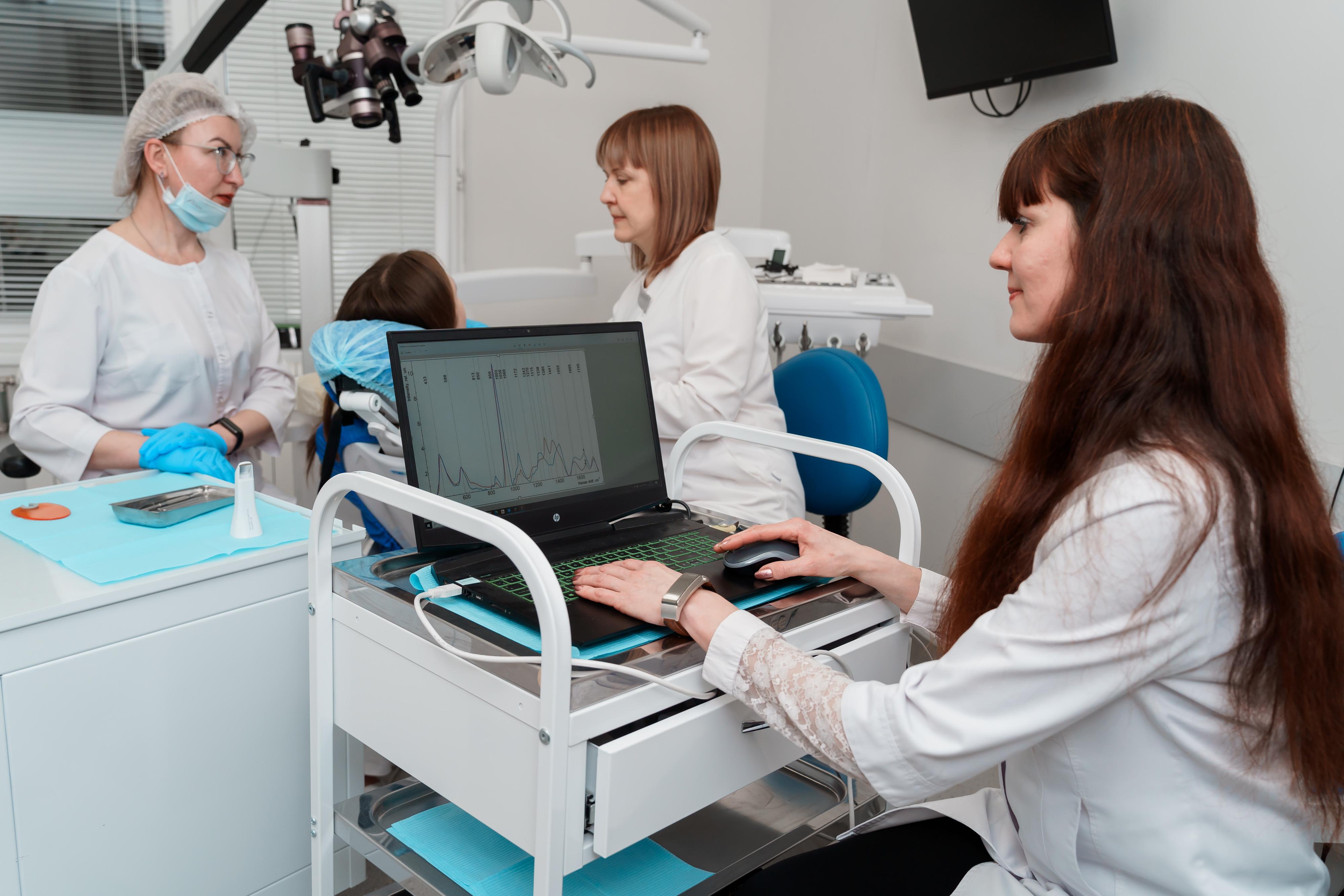
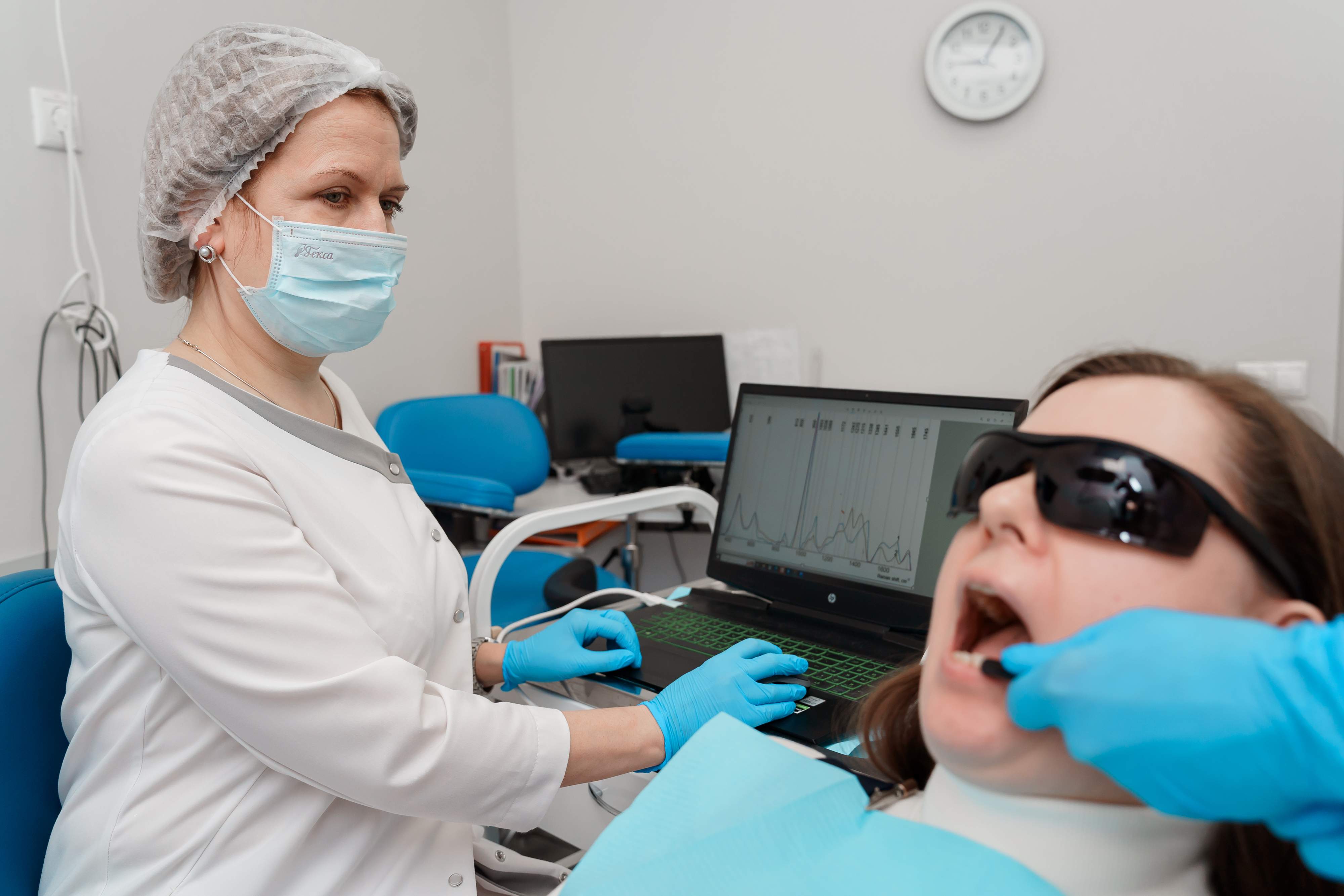
The method developed by Samara scientists improves and complements classical diagnostic methods, reducing the probability of medical error and automating the process of the disease detection. The laser method is based on the principles of Raman spectroscopy*, which allows «seeing" the spectral signs of the presence of a substance when the laser beam interacts with the object under study, in this case, with tooth enamel, by using special equipment - a spectrograph. This resembles checking a bill in a bank in the ultraviolet rays: you can see signs of protection that are invisible in normal light.
As proven in course of the studies, with periodontitis or at risk of its development, the concentration and ratio of certain substances – for example, hydroxyproline, hydroxyapatites, amide structures of types I-III – in tooth enamel are changed. This is due to the fact that diseased tissues nourish the teeth less with the necessary substances, which results in the tooth enamel to be changed, and the method allows recording this. The software program created by the scientists receives enamel "scanning" data from the spectrograph, analyzes it using a special algorithm and automatically either issues the diagnosis or warns of the possible risk of disease, if there is a spectral “imprint” characteristic of periodontitis.
"Thus, the doctor can immediately promptly check all the patient’s teeth, not only the suspicious one, but also quite healthy in appearance, without X-rays and taking tests, and detect the disease at the earliest stage. This express-diagnostics is especially useful during medical examinations or routine check-ups. If high risks of the disease are identified, the doctor will be able to prescribe preventive therapy in advance so that the disease does not occur: when using classical diagnostic methods, the disease usually has to be treated after its detection", noted Elena Timchenko.
"Despite the fact that dentistry is one of the most high-tech areas of medicine, the issues of accurate differential diagnostics of many conditions remain open. The prognosis of any disease depends on a timely diagnosis, and in the case of periodontitis, it is about the preservation or loss of teeth with all the ensuing consequences. The indisputable value of the method is in the fact that it is possible to diagnose periodontitis by using portable equipment in any dental office, which is especially important when X-ray diagnostics are unavailable. High efficiency and accuracy will make it possible to use Raman spectroscopy as an additional method for complex differential diagnosis with other dental diseases", commented Irina Bazhutova, Associate Professor of the Department of Dentistry at Samara State Medical University’s Institute of Vocational Education, Director of the Clinic "Centre for Restorative Dentistry".
For reference:
Raman spectroscopy is a type of spectroscopy that uses the ability of the studied molecules to Raman scattering of monochromatic light (laser radiation). The spectrum of scattered radiation reveals spectral lines that are absent in the spectrum of primary (excitation) light. The number and location of the new spectral lines depend on the molecular structure of the substance under research, which enables detecting certain changes in the biological tissues.
Photo by: Olesya Orina