Irina Matveyeva, a biotech engineer, Candidate of Technical Sciences, and Associate Professor at the Department of Laser and Biotechnical Systems of Samara National Research University, specializes in intelligent analysis of biomedical data. Together with colleagues from the university—and in close collaboration with Samara State Medical University (SamSMU)—she has co-developed a non-invasive, multimodal method for diagnosing skin lesions. The technique combines laser spectroscopy and high-resolution imaging, with artificial intelligence delivering rapid, accurate assessments of whether a mole is benign or malignant.
- How did this idea come about?
“This concept originated with my academic advisor, Dr. Valery Pavlovich Zakharov, Doctor of Physical and Mathematical Sciences and Professor. My senior colleagues had long been exploring various optical methods for skin disease diagnostics. Each approach reveals unique, invisible-to-the-eye information about skin lesions. Spectroscopy allows us to ‘look inside’ the skin and analyze its biochemistry, while dermoscopic imaging captures external morphological features. So we asked a logical question: Could diagnostic accuracy improve if we combined multiple optical methods—analyzing several data types simultaneously? Would such a device truly enhance clinical practice?
That became the focus of my PhD research. I successfully defended my dissertation on this topic, published several papers, and confirmed our core hypothesis: joint analysis of spectral data and dermoscopic images yields significantly better results than either method alone.
Development of the hardware—a device integrating both analytical modalities—began even before my involvement. From initial concept to today’s promising results, more than a decade has passed. Medical innovation is inherently slow, especially when it involves collecting high-quality data to train AI. Challenges include the rarity of certain conditions, diverse patient phototypes and health statuses, and strict legal and ethical constraints.”
- How does it work?
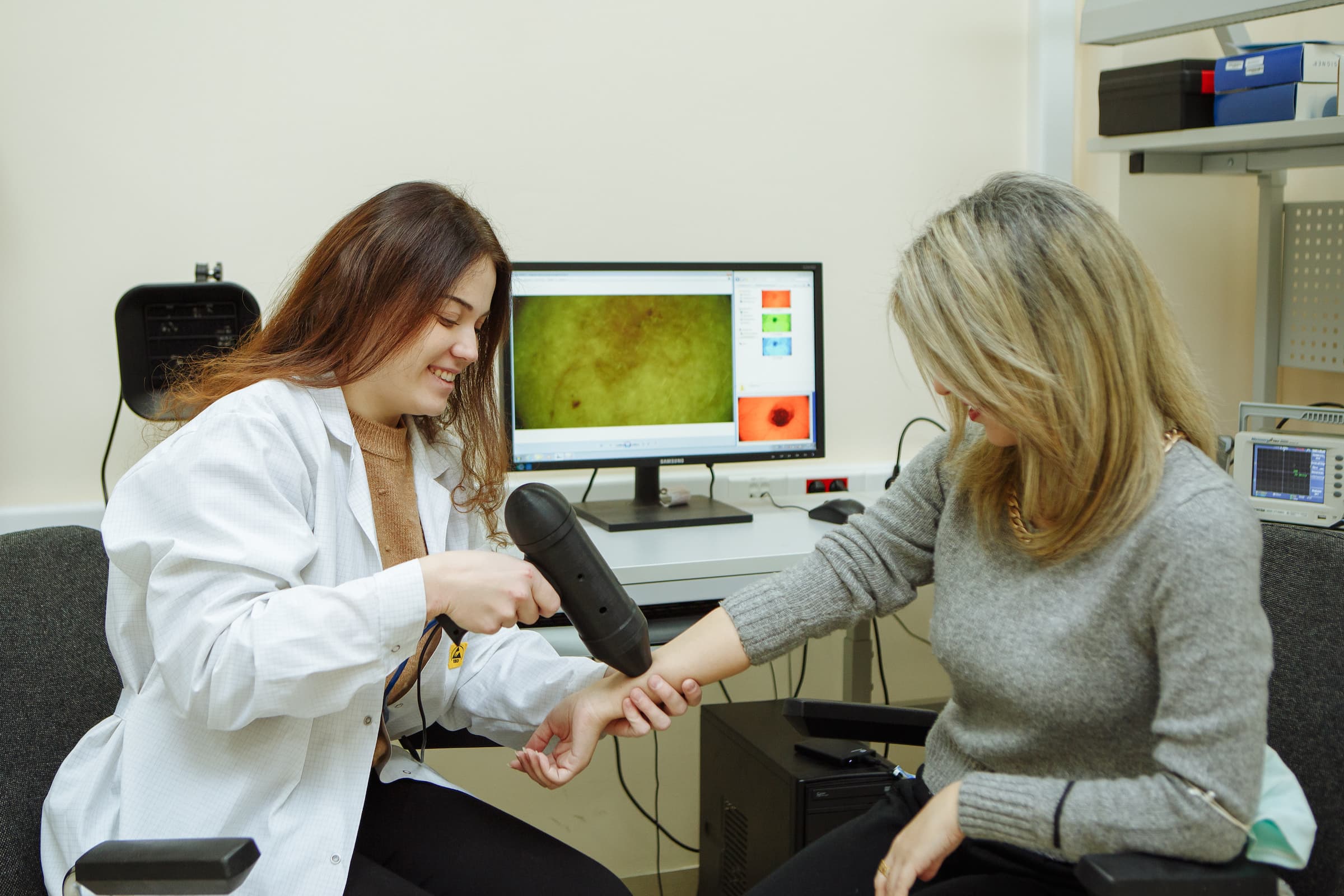
“The device has two components. The first is a Raman spectrometer—a compact box connected to a computer, containing a laser and a light detector. Laser light is delivered to the skin via optical fiber and scattered light is collected back into the instrument.
The second part is a dermatoscope, resembling a handheld camera with built-in illumination. We point it at the skin lesion to capture a high-resolution image.
First, the laser illuminates the mole and records its molecular spectrum. Then, the dermatoscope photographs it. A neural network analyzes both data streams and instantly determines whether the lesion is benign—or potentially cancerous, requiring urgent oncologist consultation.
This method could be deployed in ordinary outpatient clinics lacking specialized oncologists, as well as during routine medical screenings where rapid triage is essential: Is the patient healthy, or do they need further testing?
Our multimodal approach already outperforms visual examination by experienced oncologists. So far, we’ve trained the system to detect malignancy and specifically identify melanoma among malignant cases. But many other skin conditions also require diagnosis. To expand capabilities, we need a database of several thousand patient cases to teach the AI to distinguish between diverse pathologies. We’re now scaling up—training the system to recognize a broader spectrum of diseases.”
- Why will this invention improve people’s lives?
“Our method is completely non-invasive—no skin puncture, no biopsy. This painless, stress-free procedure is far more comfortable for patients psychologically. If implemented widely, it could enable mass screening, allowing us to detect skin cancer at earlier stages across a much larger population in our region.
Fact: Dermoscopy (even with AI) and Raman spectroscopy are well-established individually. But combining them into a single diagnostic workflow is an original idea pioneered at Samara University—no similar research exists globally.
The device is expected to cost several million rubles—but it will pay for itself quickly. It requires no consumables, and crucially, lowers staffing requirements: a general practitioner in any clinic could operate it.
Clinical trials are planned at the Samara Regional Oncology Dispensary.”
- How did you come to science?
“I was born in the small town of Pokhvistnevo in Samara Oblast and attended School No. 1. My mother is a math teacher; my father, an engineer. I’ve always been fascinated by medicine—but never wanted to become a doctor. Instead, I chose to become an engineer applying cutting-edge technologies to healthcare. That aspiration led me to Samara University’s Department of Laser and Biotechnical Systems, which offers programs in Biotechnical Systems and Technologies and Laser Engineering and Laser Technologies—both deeply rooted in medical optics and engineering. That’s how I became a biotech engineer.”
- Why is science your calling?
“I love the freedom to act, the ability to plan independently, and the constant pursuit of new knowledge. As a scientist, you choose your own direction and lead your own projects. When you make even a tiny discovery—one that might someday improve human lives—it brings profound satisfaction. I’m leaving behind something useful, meaningful, and valuable—something future generations will use. In a way, I’ll continue to ‘live’ through my inventions.”
- Who inspires you?
“I deeply admire Giordano Bruno, the Italian Renaissance philosopher, scientist, and priest. He was among the first to champion heliocentrism—and refused to recant his beliefs even at the stake of the Roman Inquisition. To me, he embodies extraordinary courage. In his time, daring to ask questions that shook centuries-old worldviews required immense bravery. Only truly great individuals defend truth and intellectual freedom at the cost of their own lives. We must look to such figures as our guiding stars.”
Source: samara.aif.ru